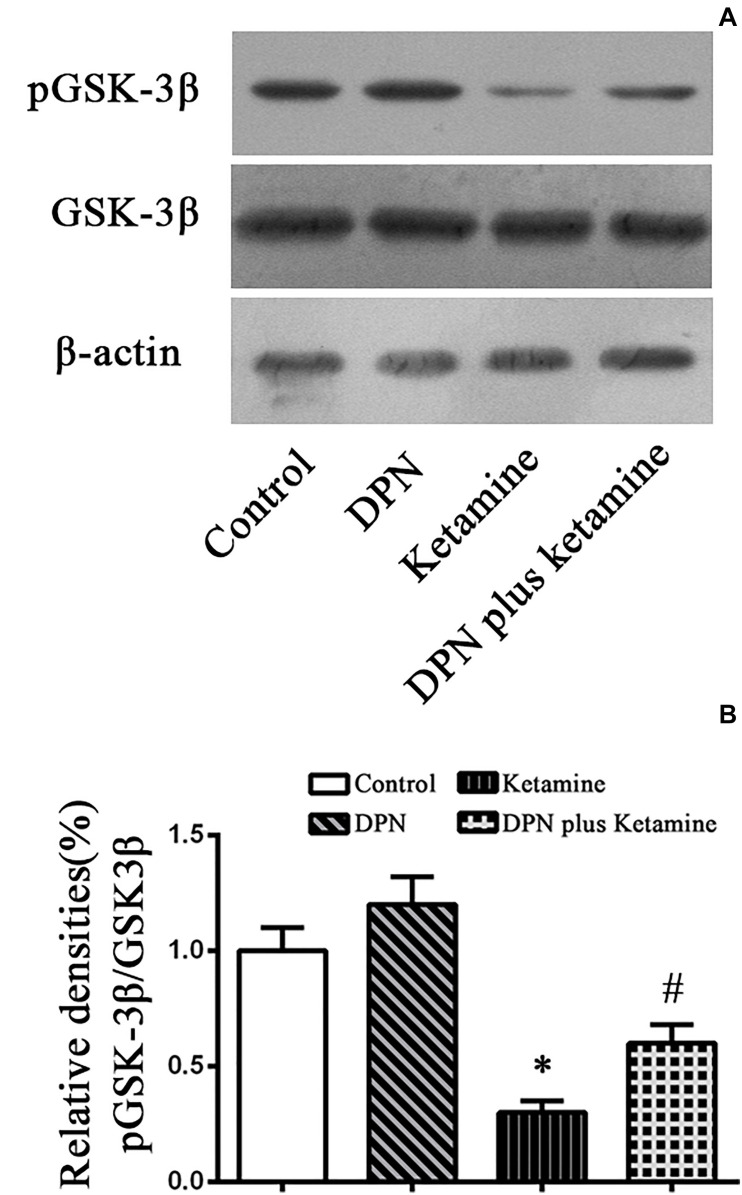
FIGURE 4.
Detection pGSK-3β protein by western blotting. (A) The typical image of Western blotting to assess pGSK-3β in NSPCs 24 h after different treatment. (B) Quantification of pGSK-3β protein level normalized to GSK-3β after different treatments. ∗P < 0.01 compared with the control, #P < 0.01 compared with ketamine group. Data were collected from three independent experiments and are presented as means ± SEM.

