Fig. 2.

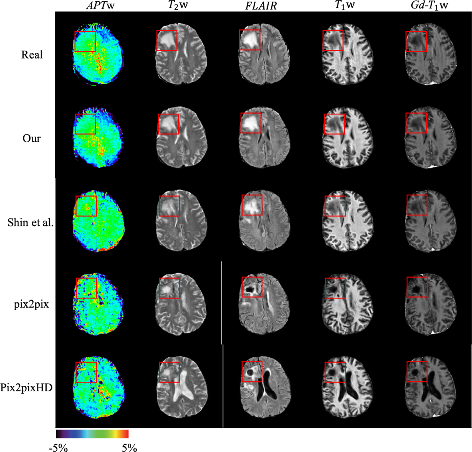
Qualitative comparison of different methods. The same lesion mask is used to synthesize images from different methods. Red boxes indicate the lesion region. (Color figure online)

Qualitative comparison of different methods. The same lesion mask is used to synthesize images from different methods. Red boxes indicate the lesion region. (Color figure online)