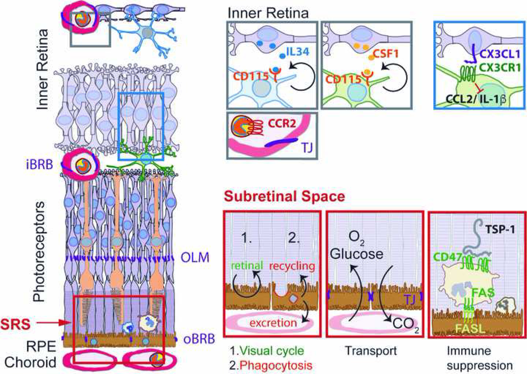
Figure 1: Physiological distribution of retinal MNPs and main functions of the subretinal space:

Physiologically the retina is only populated by IL34-dependent and CSF-1-dependent microglial cells. CCR2-positive Mos are restricted to the circulation separated from the retina by the inner and outer blood retinal barrier. CX3CL1 is constitutively expressed as a transmembrane protein in inner retinal neurons and provides a tonic inhibitory signal to the microglial checkpoint factor CX3CR1, expressed by retinal MCs. The subretinal space, demarcated by the tight junctions of the outer limiting membrane and of the RPE is the site of 1.) the visual cycle; 2.) phagocytosis of used photoreceptor outer segments and excretion or recycling the material; 3.) Oxygen and glucose uptake and CO2 excretion; 4.) immune suppression, in which FasL induces elimination of infiltrating leukocytes after activation of the integrin associated protein CD47 by thrombospondin 1 (TSP-1). iBRB: inner blood brain barrier; oBRB: outer blood brain barrier; OLM: outer limiting membrane; SRS: subretinal space; RPE: retinal pigment epithelium; IL: interleukin; CSF: colony stimulating factor; TJ; tight junction.
