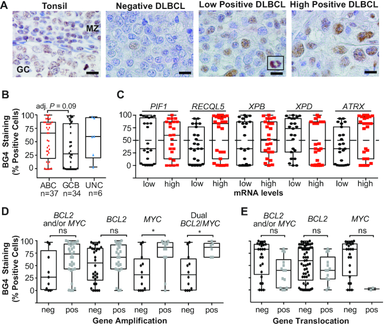
Figure 1.

BG4 staining indicates G4 formation is frequent in DLBCL tissues, particularly in non-GCB subtypes, and correlates with BCL2 and MYC amplification. (A) Representative BG4 staining in a non-malignant tonsil biopsy with positive staining within a normal GC and the MZ, and DLBCL tissues that stained negative, low or highly positive with intense staining in mitotic nuclei (inset). Original magnification is ×1000. Scale bar represents 10 μm. (B) Percent BG4-positive DLBCL cells in DLBCL tissues (n = 77) according to COO. (C) Percent BG4-positive DLBCL cells according to low or high mRNA of the given DNA helicase. Percent DLBCL cells positive for BG4 staining in tumors with BCL2 and/or MYC amplification (D) or translocation (E). Box and whisker plot: line is the median; error bars are the minimum and maximum. *Adjusted P < 0.05 as determined by a two-tailed t-test with Šídák’s multiple test correction; ns, not significant.
