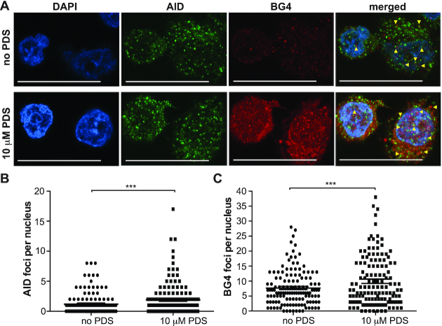
Figure 4.
AID localizes to G4s in DLBCL cell nuclei. (A) Immunofluorescence for AID and BG4 in DLBCL cells without (top panel) and with (bottom panel) PDS treatment (10 μM for 2 min). Discrete AID (green) and BG4 (red) were observed in the nucleus. Nuclei were counterstained with DAPI (blue). Yellow arrowheads indicate co-localization. Scale bars represent 20 μm. (B) Quantification of AID foci number per nucleus in cells without and with PDS treatment. (C) Quantification of BG4 foci number per nucleus without and with PDS treatment. One hundred to one hundred fifty nuclei were counted and the SEM was calculated from three independent experiments. ***P < 0.001 as determined by a Poisson exact test.

