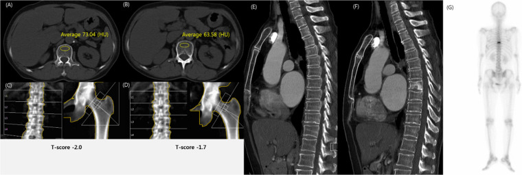
Fig 4. Compression fracture in T8 vertebral body in a 61-year-old woman with breast cancer.
(A-B) Chest computed tomography (CT) scan showed a markedly decreased L1 trabecular attenuation in 2013 (73 HU, A) and 2014 (64 HU, B). (C-D) Dual-energy X-ray absorptiometry (DXA) was interpreted as osteopenia with a T-score of -2.0 in 2013 (C) and a T-score of -1.7 in 2014 (D). (E-G) Chest CT scan showed no fracture in 2013 (E). Sagittal CT scan (F) in 2014 and bone scan (G) showed a compression fracture in the T8 vertebral body. The diagnosis discrepancy between DXA and CT in this case suggests that DXA was falsely negative given the subsequently-identified fracture.

