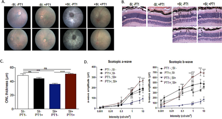
Fig 7. PT1 attenuates retinal degeneration in a mouse model of mitochondrial dysfunction.
Sodium iodate-treated (+SI) or untreated (-SI) mice were treated with PT1 or DMSO (vehicle). The four groups (n = 5 mice per group) were evaluated by (A) fundoscopy; (B) H&E staining of retinal sections showing the outer nuclear layer (ONL; white bars), pigmented cells (black arrows), and photoreceptor inner outer segment (IS/OS) morphology; (C) quantitative assessment of ONL thickness; and (D) functional evaluation by ERG with measurements of the dark-adapted photoreceptor response (scotopic a-wave) and inner retinal response (scotopic b-wave). ERG data represent 3–5 mice per group and are plotted as mean ± SEM. *P<0.05; **P<0.01; ***P<0.001; ****P<0.0001.

