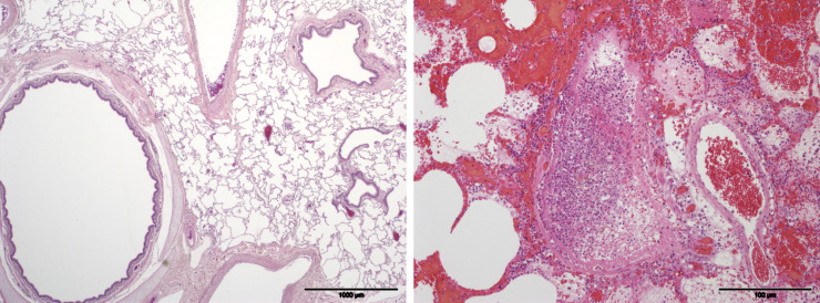
Fig 1.
Example pulmonary histopathology slides from a study 1 pig given saline by gavage (A) and a study 2 pig given organophosphorus insecticide by gavage then placed in a lung (B). The lung parenchyma and airways in Fig A are within normal limits. In Fig B most of the alveolar spaces are filled with blood, edema fluid and moderate to large numbers of neutrophils. The structure in the centre is a bronchiole filled with neutrophils admixed with fibrin. The bronchiolar epithelial lining is virtually completely necrotic and sloughed. Porcine lung, HE stained.

