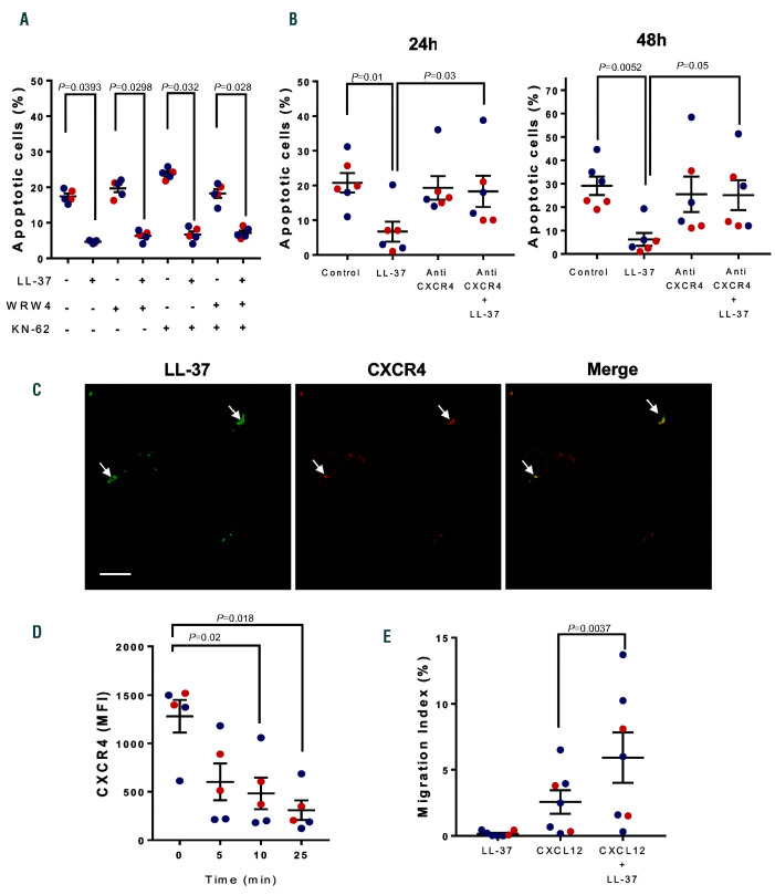
Figure 3.
Role of CXCR4 in LL-37 effects on chronic lymphocytic leukemia cells. Peripheral blood mononuclear cells (PBMC) from chronic lymphocytic leukemia (CLL) patients were incubated with WRW4 (1 mM), KN-62 (1 mM) or both for 30 min before adding LL-37 (5 mM). Cells were cultured for 48 hours (h) at 37ºC and apoptosis was evaluated as described in Figure 2. (A) The percentages of apoptotic CLL cells are shown (mean ± standard error of the mean [SEM], n= 5). Statistical analysis was performed using Friedman test and Dunn’s multiple comparison test (P<0.05). (B) PBMC from CLL patients were incubated with anti- CXCR4 Ab (10 mg/mL) for 30 min before adding LL-37 5 mM. Cells were cultured for 48 h at 37ºC. A fresh aliquot of anti-CXCR4 was re-added at 24 h. Apoptosis was evaluated at 24 and 48 h as described in Figure 2. Shown are the percentages of apoptotic cells (mean ± SEM, n=6). Statistical analysis was performed using Friedman test and Dunn’s multiple comparison test (P<0.05). (C) CLL cells were incubated for 30 min with LL-37 (5 mM), then fixed with 4% paraformaldehyde and labeled with rabbit anti-LL-37 IgG followed by Dy-Light 488-anti-rabbit IgG (green) and anti-CXCR4-PE IgG (red). Colocalization areas are indicated by the arrowheads. The bar indicates 15 mm. (D) Time-dependent downregulation of CXCR4 on CLL cells induced by LL-37. CLL cells were incubated with LL-37 (5 mM) at 37ºC for the indicated time and the expression of membrane CXCR4 was evaluated by flow cytometry. Results are expressed as mean flouresence intensisty (MFI) (mean ± SEM, n=5). Statistical analysis was performed using Friedman test and Dunn’s multiple comparison test (P<0.05). (E) LL-37 enhances the migration of LLC cells towards CXCL12. CLL cells (2x10⁶ cells/mL) were seeded in the upper chamber of transwell plates to evaluate their migration to the lower compartment in response to CXCL12 (25 ng/mL) in the presence or absence of LL-37 (5 mM). Cells were incubated for 120 min, recovered from the lower chamber and quantified by flow cytometry. The migration index was calculated as the subtraction of the number of CD19+ cells that migrated spontaneously (control without chemokine) from the number of CD19+ cells that migrated in the presence of CXCL12 and normalized to the input. Shown are mean ± SEM (n=7). Statistical analysis was performed using Friedman test and Dunn’s multiple comparison test (P<0.05). In all cases red dots represent U-CLL and blue dots M-CLL.

