Figure 1.
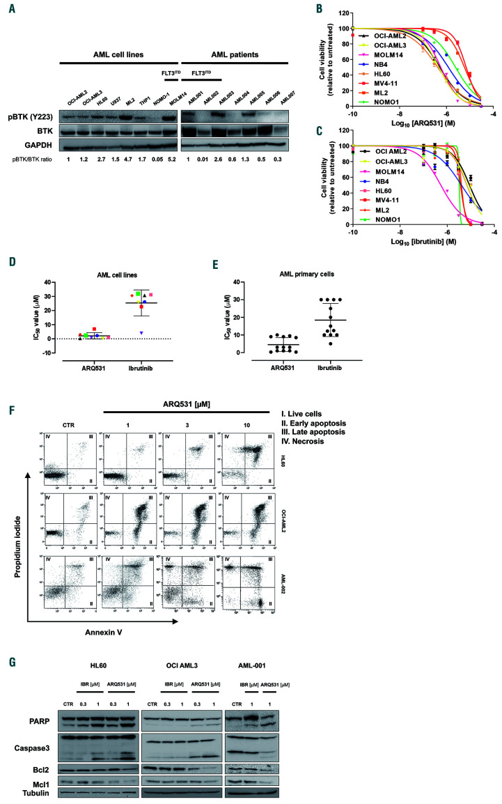
ARQ531 shows s trong anti-tumor activity by inducing apoptosis of acute myeloid leukemia cells. (A) Immunoblot for phospho- BTK, BTK and GAPDH (loading control) in the indicated human acute myeloid leukemia (AML) cell lines and primary AML samples regardless of the specific g enomic lands cape. (B, C) Viability of AML cell lines after treatment with ARQ531 (B) or ibrutinib (C), as measured by MTS assay. The mean ± standard deviation (SD) from at lea st three indepe ndent exp eriments is shown. (D) Half maximal inhibitory concentration (IC50) values, measured for each tested cell line as in (B) and (C). (E) Drug effects on primary AML patie nt-derived samples (n=13 ) treated with increasing doses of ARQ531 or ibrutinib (0-30 mM for 48 h). IC50 values are visualized for each tested primary AML cell line. (F) HL60, OCI-AML2 and primary AML-002 cells were treated with ARQ531 or dimethylsulfoxide (CTR) in a dosedependent manner for 48 h. Apoptotic cells were detected by annexin V/propidium iodide staining. Representative dot plots are shown. (G) Immunoblots for PARP, caspase 3, MCL-1, BCL-2 and tubulin on indicated AML cell lines and primary blast cells following treatment with a BTK inhibitor (ARQ531 versus ibrutinib) at 24 h.

