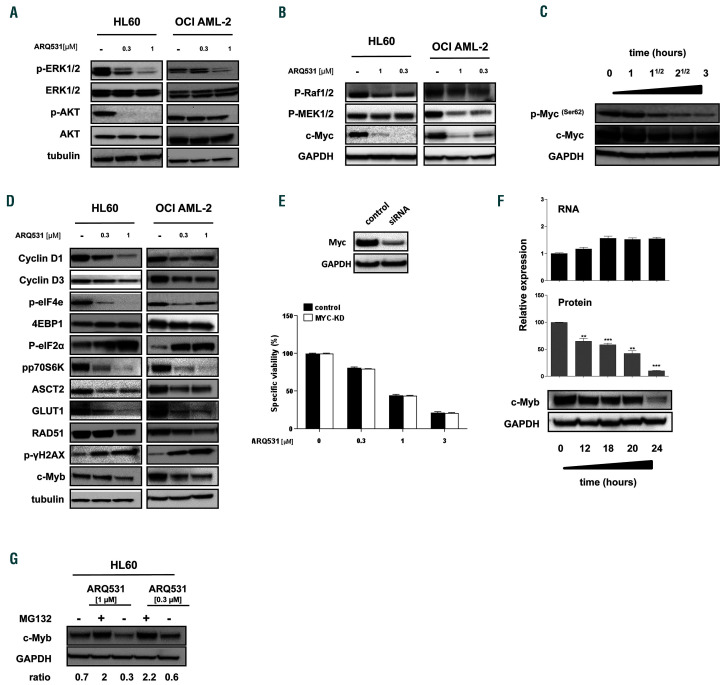
Figure 4.
BTK inhibition and MYC/MYB degradation represent molecular bases for the anti-leukemic activity of ARQ531. (A) Western blot showing that 24 h of treatment with ARQ531 (0.3-1 mM) abrogates ERK and AKT activation in HL60 and OCI-AML2 cells. (B) Western blot analysis shows that 24 h of ARQ531 treatment (0.3-1 mM) affects kinases in the RAF/MEK/ERK pathway of acute myeloid leukemia (AML) cell lines, resulting in MYC downregulation. (C) Western blot showing time-dependent ef fects of ARQ531 exposure on p-MYC S62 and total MYC in the HL60 cell line. (D) Western blot showing deregulation of c-MYCcontrolled signals in AML cells following treatment with ARQ531 at the indicated doses after 24 h. (E) Viability of MYC-silenced or control HL60 cells treated with increasing doses of ARQ531 for 48 h. The mean ± standard deviation (SD) are shown (n=3). (F) Protein and mRNA expression in AML cells af ter 24 h treatment with dimethylsulfoxide (DMSO) or the indicated ARQ531 concentrations, normalized to DMSO controls. Bars and error bars are means and SD of three independent experiments. *P<0.05; **P=0.01; ***P<0.001; n.s. not significant (relative to DMSO controls), one sample t-test. Western blots below graphs show examples of MYB protein expression. (G) Western blot analysis of MYB protein expression in AML cells after 24 h treatment with DMSO, ARQ531 (0.3-1 mM) or ARQ531 and 10 mM MG132, a proteasome inhibitor.

