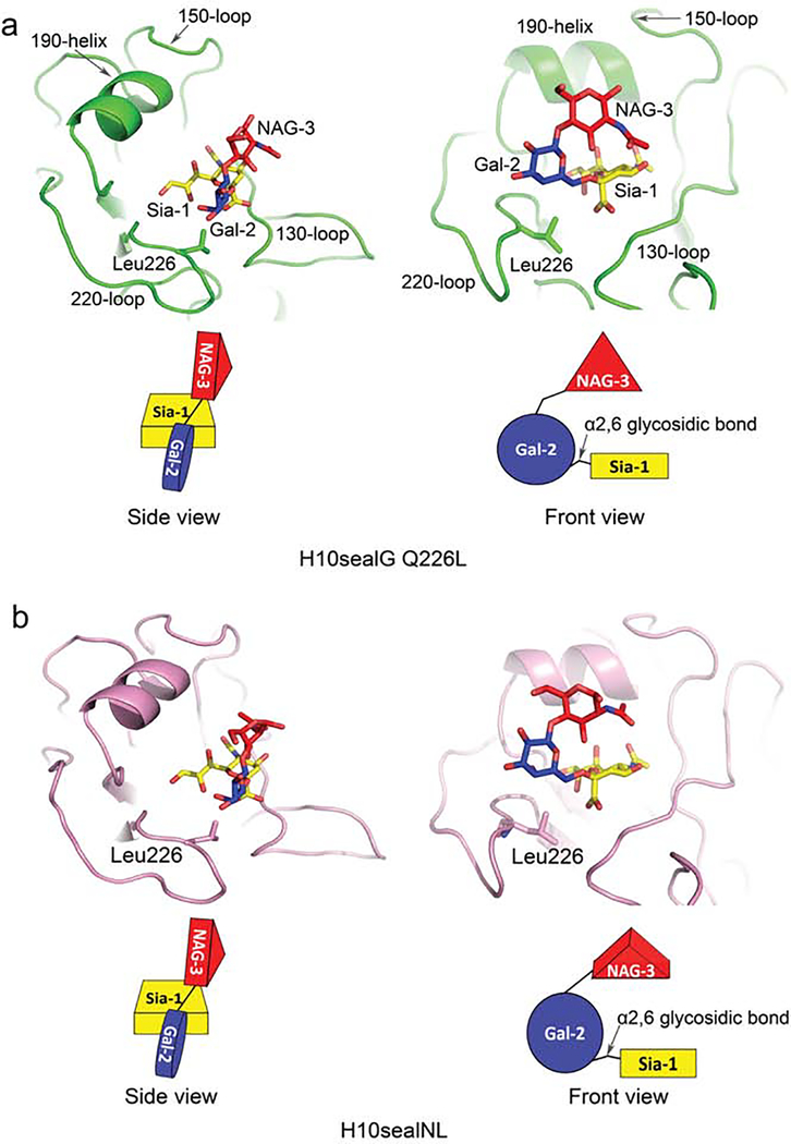
Figure 5.
Human-type receptor binding features of H10sealG Q226L and H10sealNL. The side and front views of conformations of human-type receptors bound to H10sealG Q226L and H10sealNL are shown on the left and right sides of panel (a-b) respectively. The schematic representations of the conformations of the human-type receptors are shown in panel (a-b) as well. The sialic acid, galactose and N-acetylglucosamine of receptor analogue are coloured in yellow, blue and red individually. H10sealG Q226L is coloured in green in its complex structure (a). HAsealNL is coloured in pink (b).

