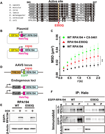
Fig. 3. Mutant Pol I with an RPA194 variant (RPA194-E593Q) that causes a craniofacial disorder.

(A) Amino acid sequence comparison of RPA194 including the active site for Mg2+ binding among Homo sapiens, Mus musculus, Danio rerio, Xenopus laevis, Drosophila melanogaster, Saccharomyces cerevisiae, and Escherichia coli. In the RPA194 variant from the patient, the conserved glutamic acid residue (E) at position 593 (highlighted orange) was substituted with glutamine (Q). (B) Diagram of plasmids expressing HaloTag-WT RPA194 and HaloTag-RPA194-E593Q used for single-molecule imaging. (C) MSD plots of HaloTag-RPA194 molecules after expressing HaloTag-RPA194 (WT, black, n = 18 cells) or HaloTag-RPA194-E593Q (E593Q, red, n = 17 cells), with 95% CIs. For comparison, MSD data for WT HaloTag-RPA194 with CX-5461 treatment are reproduced from Fig. 2D (red). **P < 0.01 via bootstrapping for the WT RPA194 versus RPA194-E593Q (P = 5.2 × 10⁻³). (D) Diagram of the clone 2 cell line (Fig. 1B) expressing EGFP-RPA194-E593Q (or EGFP-RPA194) from an exogenous AAVS locus. (E) Expression of EGFP-RPA194-E593Q induced by doxycycline (DOX; 2 μg/ml) for 24 hours. (F) Coimmunoprecipitation of endogenous HaloTag-RPA194 with EGFP-RPA194 or EGFP-RPA194-E593Q. I, input; N, negative control precipitant (HaloTag-ligand minus); P, precipitant (HaloTag-ligand plus).
