. 2020 Sep 10;9:e52253. doi: 10.7554/eLife.52253

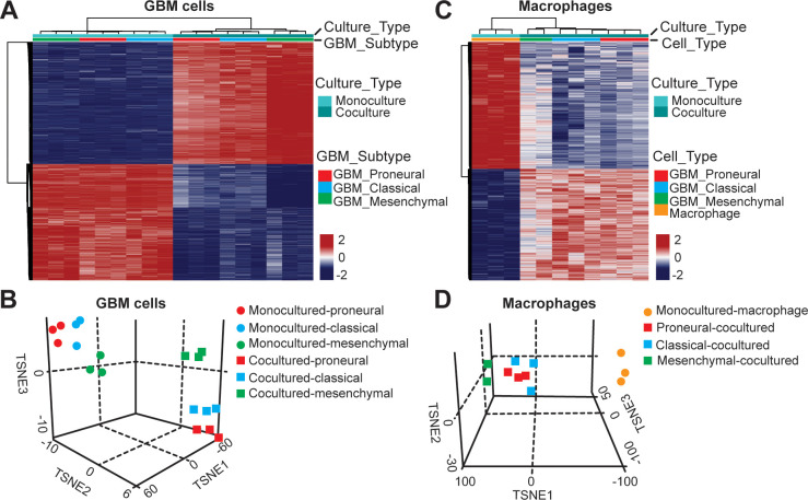
Figure 4. DNA methylation analysis of interactions between patient-derived GBM cell and macrophage in an engineered 3D GBM niche environment.

(A) Whole genome DNA methylation analysis showing top 10,000 differentially methylated probes of patient-derived PN (GBML20), CL (GBML08) and MES (GBML91) GBM cells cultured in a 3D brain-mimicking ECM environment with or without macrophages. (B) tSNE analysis of mono-cultured and co-cultured GBM cells showing clear separation of all molecular GBM subtypes, PN (GBML20), CL (GBML08) and MES (GBML91) (each in triplicate) in the same direction when exposed to macrophages. However, the effect appears to be different in the three molecular subtypes with PN GBM mostly affected by presence of macrophage. (C) Whole genome DNA methylation analysis showing top 10,000 differentially methylated probes of mono-cultured and GBM cell-educated macrophages. (D) tSNE analysis of mono-cultured and patient-derived GBM cell-educated macrophages showing distinct shifts in methylation in all molecular GBM subtypes. However, MES GBM cell co-cultured macrophages cluster showed a more distinct separation.

Figure 4.

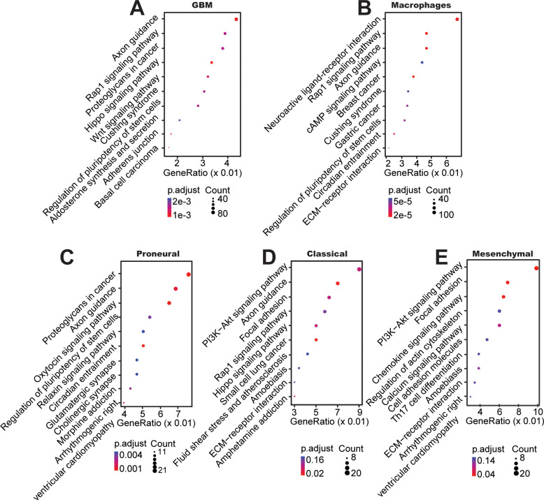
Figure 4—figure supplement 1. Top KEGG pathways between mono-cultured and co-cultured GBM cells in a 3D brain-mimicking ECM environment.

Figure 4—figure supplement 1.

(A) KEGG pathway analysis from whole genome DNA methylation comparing data from all glioma subtypes co-cultured with macrophages vs all mono-cultured glioma subtypes including PN (GBML20), CL (GBML08), and MES (GBML91) showing activation of pathways involved in axon guidance, Rap1, proteoglycans, and Wnt related signaling. (B) KEGG pathway analysis of macrophages co-cultured with all different types of GBM cells compared with mono-cultured macrophages showed relatively activation of pathways in neuroactive ligand-receptor, Rap1, axon guidance, cAMP related signaling. When DNA methylation data were analyzed for each molecular subtype of GBM, (C) co-cultured PN (GBML20) GBM cells showed relatively higher ratios of genes in proteoglycans, axon guidance, oxytocin, and pluripotency of stem cell related signaling compared to mono-cultured GBM cells, (D) co-cultured CL (GBML08) GBM cells showed relatively higher ratios of genes in PI3K−Akt, axon guidance, focal adhesion, and Rap1-related signaling pathways compared to mono-cultured GBM cells, and (E) co-cultured MES (GBML91) GBM cells showed relatively higher ratios of genes in PI3K−Akt, focal adhesion, chemokine, and actin-related signaling pathways compared to mono-cultured GBM cells.
Figure 4—figure supplement 2. PD-L1 promoter methylation in mono-cultured and co-cultured GBM cells in a 3D brain-mimicking ECM environment.

Figure 4—figure supplement 2.

The absence or presence of macrophages does not induce changes in methylation of the PD-L1 gene promoter.
Figure 4—figure supplement 3. Analysis of extracellular matrix composition in different engineered GBM niches.
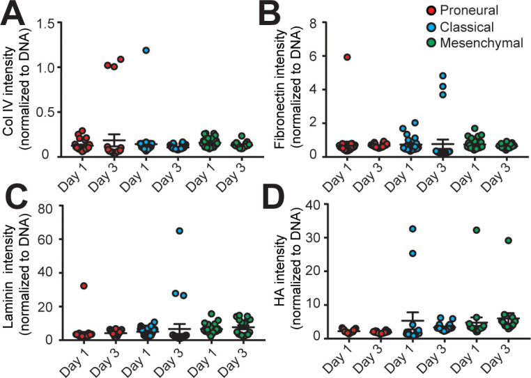
Figure 4—figure supplement 3.

The quantified results of (A) Collagen IV (Col IV), (B) Fibronectin, (C) Laminin, and (D) hyaluronic acid (HA) depositions in the MES (GBML91), CL (GBML08) and PN (GBML20) GBM niches showed no significant changes in the 3-day culture period. Error bars represent ± s.e.m.