. 2020 Oct 7;9:e56533. doi: 10.7554/eLife.56533

Figure 6. Biofilms deform and disrupt epithelial cell monolayer.

(A) CMT-93 and MDCK cells grow at the surface of a soft ECM into a tight monolayer on which we seed a liquid inoculum of V. cholerae Rg. (B) Confocal images of uninfected (i) and infected (ii-v) monolayers of CMT-93 cells. Yellow arrow indicates gaps in the epithelial monolayer (ii and iii), blue arrow shows deformed ECM (iv). (C) Confocal images of uninfected (i) and infected (ii-iii) monolayers of MDCK cells, also showing delamination and rupture as illustrated in (D). Scale bars: 20 µm.

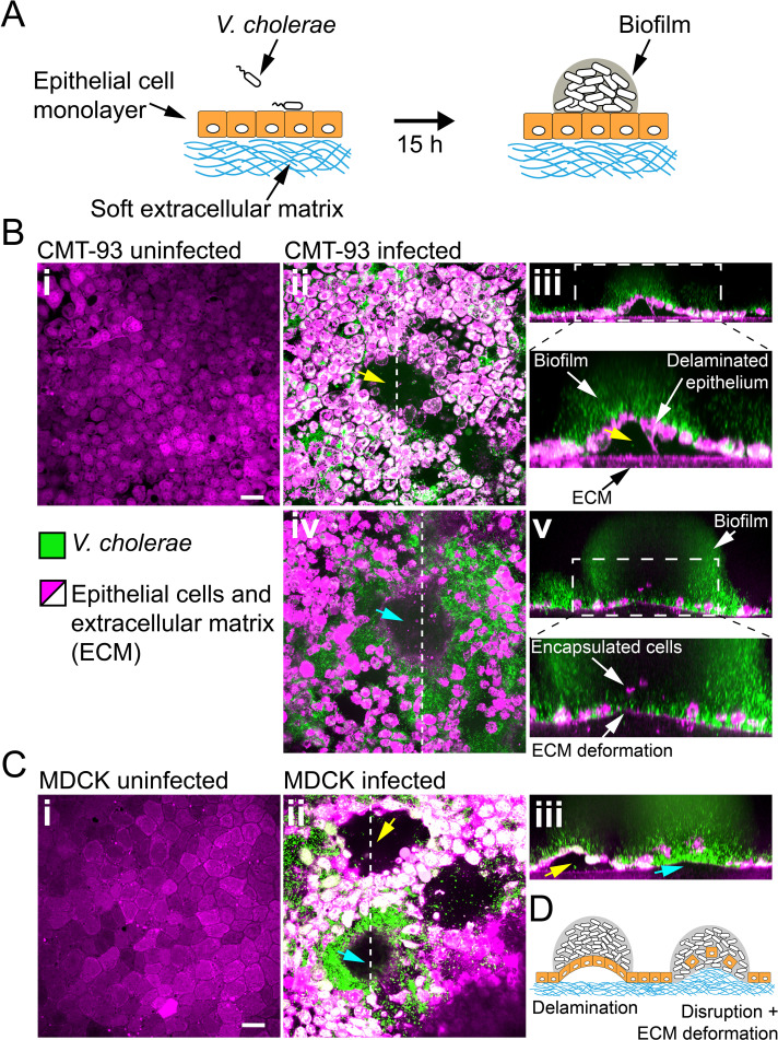
Figure 6.

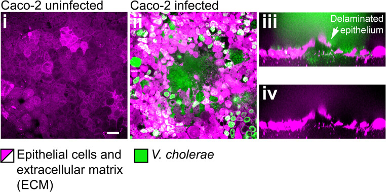
Figure 6—figure supplement 1. Confocal images of uninfected (i) and infected (ii-iv) monolayers of Caco-2 cells.

Figure 6—figure supplement 1.

Scale bars: 20 µm.
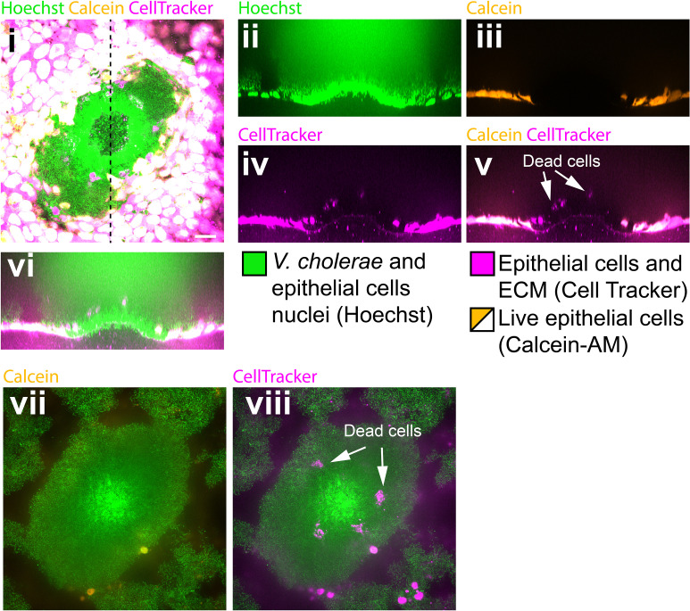
Figure 6—figure supplement 2. Biofilms perturb the viability of MDCK cell monalyers.

Figure 6—figure supplement 2.

(i) Confocal in plane visualization of a Vc Rg biofilm growing on top of an MDCK cell monolayer stained with CellTracker, Hoechst and Calcein-AM. Cross-section visualization of the infected MDCK monolayer stained with Hoechst (ii), Calcein-AM (iii), Cell Tracker (iv) and merged visualizations (v, vi). (vii, viii) Confocal in plane visualization of the biofilm on a focal plane above (i). Scale bars: 20 µm.
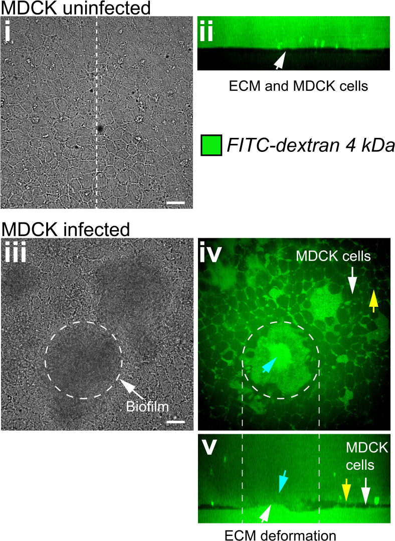
Figure 6—figure supplement 3. Biofilms increase the permeability of MDCK cell monolayers.

Figure 6—figure supplement 3.

(i) Bright-field in plane image of an uninfected MDCK cell monolayer grown at the surface of a soft ECM. (ii) Confocal cross-section shows that uninfected monolayers are impermeable to FITC labeled Dextran. (iii) Bright-field in plane visualization of an MDCK cell monolayer infected with Vc Rg. The dashed line shows the approximate edge of the biofilm. Confocal in plane (iv) and cross section (v) visualization of FITC-labeled Dextran permeability through the damaged epithelium. Blue arrows show Dextran permeability through the biofilm in direct contact with the ECM, yellow arrows indicate Dextran permeability through epithelial cells junctions. Scale bars: 20 µm.