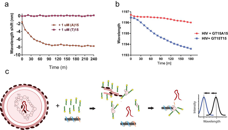
Figure 5:
Detection of intact virus RNA using a HIV lentivirus model. a. Kinetics of wavelength shift of nanotube suspended with the DNA oligonucleotide (GT)15-(T)15, focusing on the (8,6) nanotube species, upon introducing target oligonucleotide, (A)15, or non-target control, (T)15, with 0.2% SDBS. b. Kinetics of the response of (GT)15-(T)15 or (GT)15-(A)15-suspended nanotubes with concentrated HIV and 2% SDS. c. Model depicting HIV RNA detection. The virus is ruptured and denatured by SDS, liberating the RNA genome, which hybridizes to the sensor, freeing space on the nanotube surface. Denatured viral proteins bind to the freed space on the nanotube surface, eliciting an enhanced blue-shifting response.

