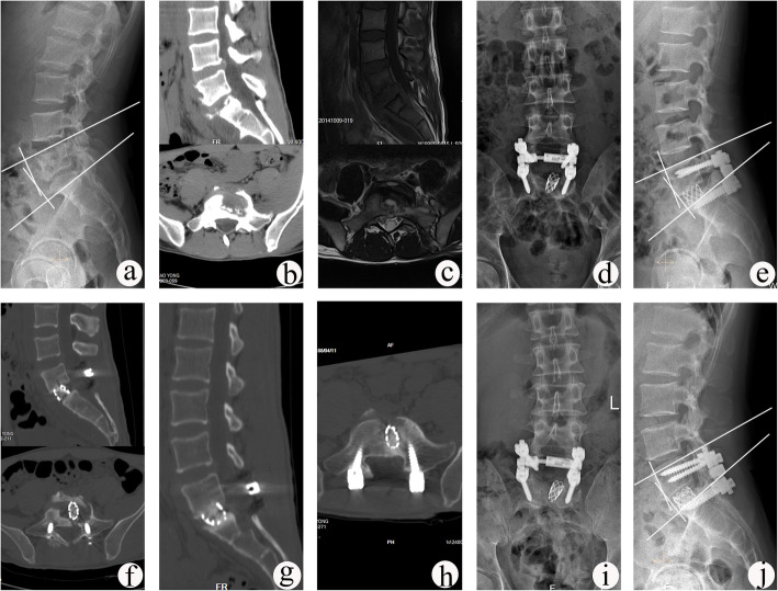
Fig. 2.
A 26-year-old male with L5–S1 TB underwent one-stage posterior debridement, titanium mesh cage bone grafting and single-segment fixation. a–c Preoperative images show lesions with lumbosacral angle of 12° and upper DH of 10.7 mm. d–f Postoperative X-ray demonstrating correction of the deformity (lumbosacral angle was 18°) and CT findings showing that the titanium mesh cage with autogenous bone particles was implanted into vertebral body. g, h CT images showing satisfactory bone fusion at 12 months after surgery. i, j X-ray displaying good internal fixation position and solid bone fusion, with the lumbosacral angle of 17° and the upper DH of 10.5 mm at the follow-up period of 81 months

