
Figure 6. Whole kidney imaging using Ultramicroscope Blaze with extended sample holding capacity.
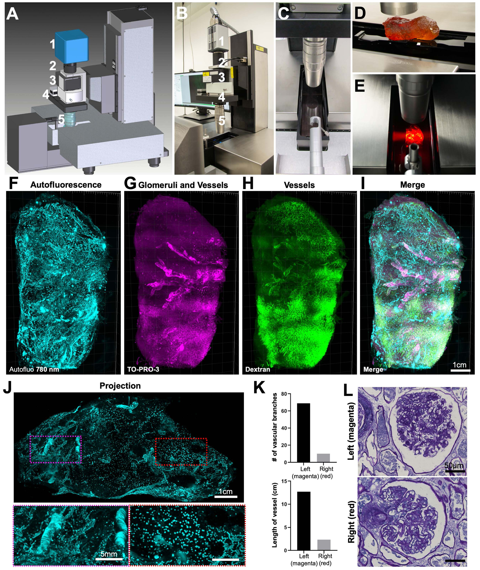
Plan (A) and picture (B) of the prototype LaVision (Miltenyi) Biotec light-sheet Ultramicroscope Blaze for large samples, featuring (1) Andor sCMOS camera, (2) tube, (3) LaVision autofocusing unit for automatic focus correction at different wavelengths, (4) zoom body, (5) 1.1x MI PLAN objective.
(C) Imaging chamber.
(D) A whole adult human kidney was mounted on the holder (note that the sample does not look transparent if not immersed in RI matching solution (BABB) as shown in the image).
(E) View of cleared whole human kidney placed in the imaging chamber with the light-sheet from the left crossing through the sample.
(F-I) 3D reconstruction of whole adult human kidney (original size of 11 × 6.5 × 5 cm) imaged by the prototype light-sheet microscope. Shown are the auto-fluorescence signal at 780 nm (F, cyan), the glomeruli and vessels from TO-PRO-3 labeling (G, magenta), the vessels from the dextran labeling (H, green), and themerged view of (F)–(H) in (I).
(J) An orthogonal 1 mm projection of the kidney showing that vascular structures are significantly reduced at the right side (red-dashed region) compared to left side of the kidney (magenta-dashed region).
See also Movie S4.
(K) Quantification of vascular features between the left (magenta) and right (red) regions of the kidney.
(L) Periodic Acid Schiff (PAS) images of rehydrated samples dissected from the left (magenta) and right (red) regions showing similar glomeruli structures for both sides.