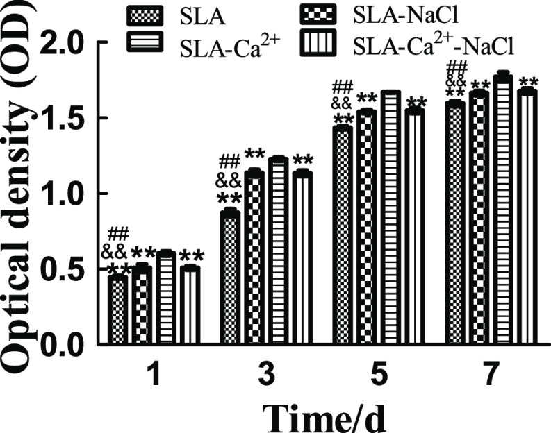
Figure 9.
Proliferation of MG63 cells seeded onto SLA, SLA-NaCl, SLA-Ca2+, and SLA-Ca2+-NaCl surfaces as measured by the MTS assay. The SLA-Ca2+ group had significantly higher proliferative activity than the other three groups, regardless of the incubation time (P < 0.01). There were no significant differences in proliferative activity between the SLA-NaCl and SLA-Ca2+-NaCl groups at the time points used in this study, but the two groups showed significantly higher proliferative activity than the SLA surface at all incubation times (P < 0.01). **P < 0.01 compared to the group of SLA-Ca2+, ##P < 0.01 compared to the group of SLA-NaCl, and &&P < 0.01 compared to the group of SLA-Ca2+-NaCl.

