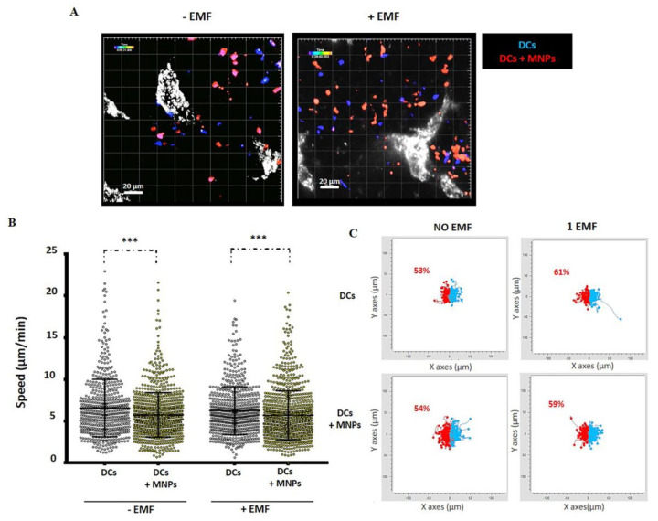
Figure 6.
In vivo behavior of MNP-loaded or unloaded murine DCs in the popliteal LN in the presence or absence of an EMF. (A) Representative captures of multiphoton microscopy movies obtained in presence or absence of EMF (blue: MNP-free DCs, red: MNP-associated DCs, gray: high endothelial venules (HEVs)). (B) Quantification of cell speed after movie analysis using Imaris software. The results shown (mean ± SD) are representative of two–three independent experiments. Student’s t-test, *** p < 0.001. (C) Paths followed by MNP-treated or untreated DCs inside the LN during the multiphoton microscopy assays in the absence or presence of EMF placed on the left (overall DC displacement to the left in red; to the right in blue), analyzed with the software provided by Ibidi (Chemotaxis and Migration tool). The methodology and ethic committee permits for these experiments are detailed in [78].

