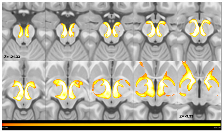
Figure 3.
Multiple axial slices showing the course of thresholded MPMs of tracts joining the PPN and GPe. MPMs have been superimposed on the ICBM template and displayed in the form of a lightbox, which shows the course of the average maps in a caudal-to-cranial direction. As shown by color bars, the voxel intensity is proportional to the number of subjects; thus, voxels characterized by lowest intensities overlapped in no more than 50 subjects, whereas voxels characterized by highest intensities overlapped in all subjects. Maps joining the PPN to the GPe ascend through the mesencephalic tegmentum in the most caudal slices and reach the GPe after entering the cerebral peduncle in the most cranial slices. Left and Right are reported according to radiological convention.

