Figure 1.
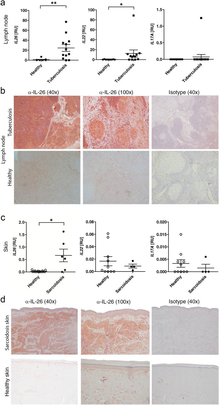
IL-26 is over-expressed in tuberculous lymph nodes and cutaneous sarcoidosis. (a, c) qPCR analysis of gene expression of IL26, IL22 and IL17A in tuberculous LN (a, n = 12) compared to healthy control LN (n = 9) in RNA from formalin-fixed paraffin-embedded (FFPE) lymph nodes and sarcoidosis skin punch biopsies (c, n = 4–6) compared to healthy skin controls (n = 10). qPCR-values are depicted as relative units compared to 18S RNA expression. Data are presented as single values and mean ± SEM. Mann–Whitney U test was used to evaluate significant differences (*p < 0.05, **p < 0.01 and ***p < 0.001). (b, d) immunohistochemistry with anti-IL-26 and isotype control on FFPE lymph node (LN) sections (10 µm) from one representative tuberculosis patient or healthy control and sections (4 µm) from skin punch biopsies from one representative sarcoidosis patient and one healthy skin donor. Magnification: ×40 for left and right panel, ×100 for middle panel.

