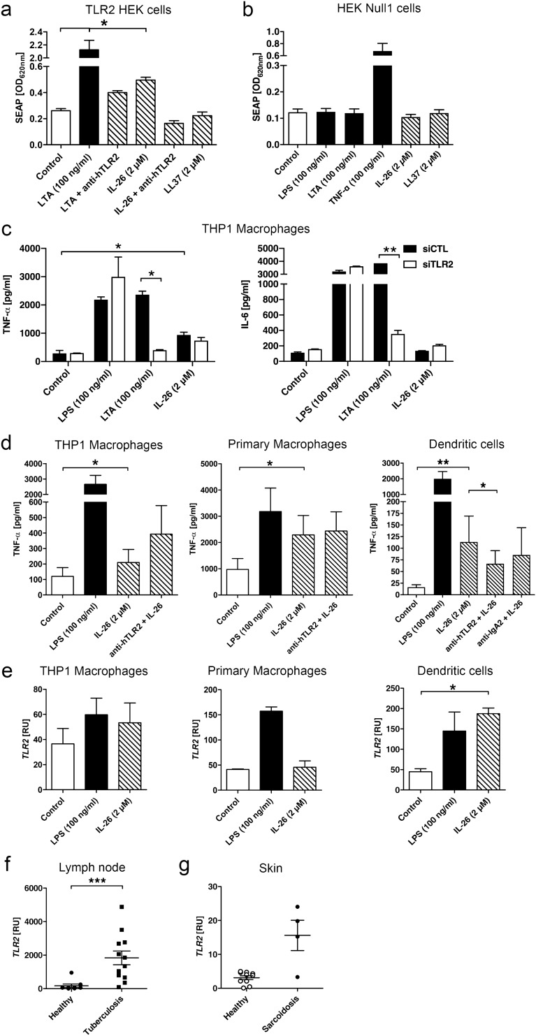
Figure 4.
IL-26 exerts effects on moDCs via TLR2. (a) Secreted embryonic alkaline phosphatase (SEAP) reporter assay was used to determine if IL-26 is able to signal via TLR2 (n = 4). (b) HEK Null1 cells are the parental cell line and are used as controls (n = 3). Statistical analysis was done using Mann Whitney U test (* equals p < 0.05). (c) TNF-α and IL-6 secretion in THP1 macrophages treated with siTLR2 or control siRNA (siCTL) (n = 2). (d) TNF-α secretion by THP1 macrophages, primary macrophages and moDCs treated with IL-26 or pretreated with anti-TLR2-antibody for 45 min before the addition of IL-26 for another 24 h and measured via ELISA (n = 6). (e) TLR2 gene expression after IL-26 stimulation in THP1 macrophages, primary macrophages and DCs (n = 5), or comparing tuberculosis (f) or sarcoidosis (g) to their respective healthy control. Data are presented as mean + SEM or a single dot is representing one independent sample. Statistical analysis was done using Wilcoxon matched pairs signed rank test (* equals p < 0.05) or Mann–Whitney U test for the diseases (*** equals p < 0.001).

