Figure 3.
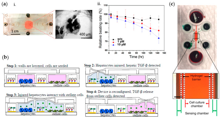
(a) Photograph of the microfluidic bioreactor and the cardiac spheroids (i); measured normalized beating rates of cardiomyocytes over time after exposure to different concentrations (0, 1, and 10 μM) of doxorubicin (ii). Adapted from Ref. [91] with permission from ACS Publications. (b) Aptamer-based two-layer device to monitor the communication between hepatocytes and stellate cells into three stages. Step 1: valves are closed to allow for cell seeding; Step 2: 100 mM EtOH in media infused into hepatocyte chamber to injure cells; Step 3: barrier raised to allow injured hepatocytes to communicate with quiescent stellate cells; Step 4: middle barrier lowered again to sequester stellate cells from injured hepatocytes and monitor TGF-β1 production from stellate cells. Adapted from Reference [92] with permission from The Royal Society of Chemistry. (c) Microfluidic system for the cultivation of hepatocytes and for the detection of secreted growth factors; photograph and microscopic image of the device; red and green food dyes were infused into the cell culture chamber and sensing chambers, respectively. Scale bar = 500 μm. Adapted from Reference [94] with permission from Springer Nature.

