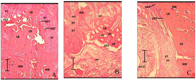
Figure 3.
Histologic findings of mandibular bone defect in groups 2 months after surgery with magnification of ×100. (A) Control (the mandibular bone defect was mainly replaced by lamellar bone), (B) low-intensity pulsed ultrasound (the mandibular bone defect was mainly replaced by lamellar and woven bone), (C) whole-body vibration (the mandibular bone defect was mainly replaced by lamellar bone). LB: lamellar bone, WB: woven bone, FT: fibrosis or connective tissue, Inf.C: inflammatory cells, BM: bone marrow, OST: osteoblast.

