Figure 2.
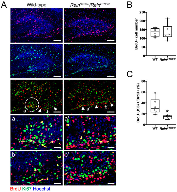
Absence of the neurogenic cluster at the fimbriodentate junction. (A) Distribution of the BrdU (red) or Ki67-positive cells (green) in the dentate gyrus at P4, after 24 h from BrdU injection. A cluster of dividing cells is present at the fimbriodentate junction of the wild-type (white circle, a). A RelnCTRdel mutant does not have this cluster (a’). Compared with a corresponding region of the wild type (b), abnormal accumulation of the dividing cells are apparent in the subpial surface of RelnCTRdel (b’). Enlarged images of marked regions (a, a’, b, b’) are shown below. In the mutant, Ki67-positive cells are closely located on top of BrdU-positive cells (a’ and b’). Scale bars, 100 µm. Scale bars in the enlarged images, 25 µm. Nuclei were stained using Hoechst (blue). (B) The number of total BrdU-positive cells is not significantly different (p = 0.8182; boxplot, median ± IQR; whiskers, min and max). (C) The percentage of BrdU/Ki67 double-positive cells was reduced (p = 0.0022; boxplot, median ± IQR; whiskers, min and max). This represents a population of cells that reentered the cell cycle.

