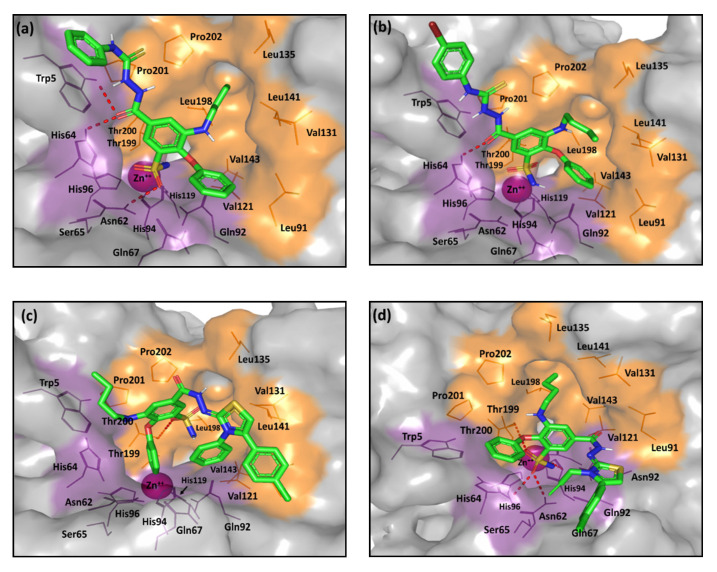
Figure 8.
The predicted binding modes of the investigated compounds (sticks) at the hCA-IX binding site (PDB ID: 3iai); (a) 7a; (b) 7c; (c) 8a; (d) 8d. The active site cleft within the suggested ligand-protein complexes is illustrated as gray surface representation depicting the prosthetic Zn(II) (magenta sphere), as well as the hydrophilic (purple) and hydrophobic (orange) residues as lines. Hydrogen bonding is depicted as red dashed-lines, while polar coordination to Zn(II) as black dashed-lines. Only residues located within 5Å radius of bound ligands are displayed (lines) and labeled with sequence number.

