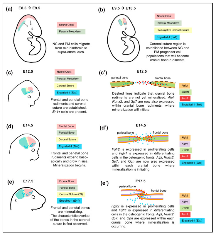
Figure 3.
Embryonic Development of the Coronal Suture with Frontal and Parietal Bones. The coronal suture exists as a physical boundary separating the neural crest-derived frontal bone and paraxial mesoderm-derived parietal bone throughout embryonic development. (a–e) Schematics depict lateral view of whole skull. (c’–e’) Schematics depict sagittal sections of the skull lateral to the midline to describe the spatial relationship of the developing frontal and parietal bones. The expression patterns of genes essential for development are depicted in various colors at indicated time points.

