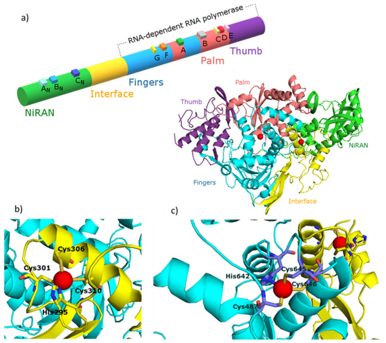
Figure 1.
(a) Diagram and structure of the SARS-CoV nsp12 protein indicating protein domains (NiRAN, interface, fingers, thumb and palm), conserved motifs (AN, BN, CN, G, F, A, B, C, D) and zinc(II) binding sites (red spheres). Enlarged zinc(II) binding sites of nsp12 protein placed in: (b) interface region and (c) fingers region. The figure was generated using PyMOL [41]. PDB entry: 7BTF [42]. Figures based on [21,22].

