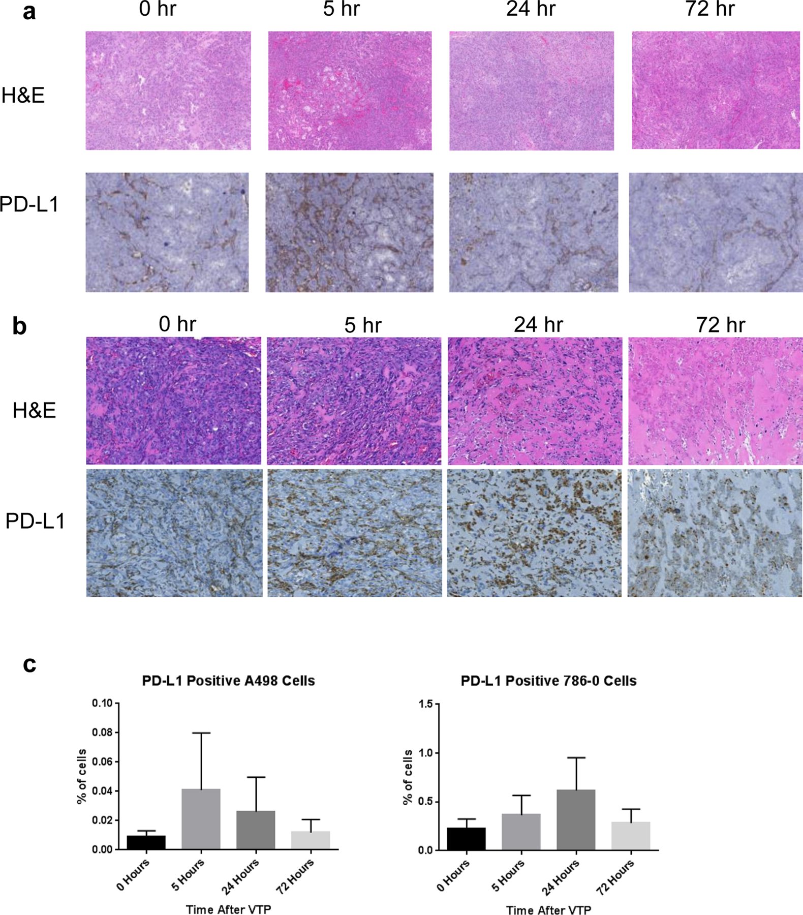
Figure 5. VTP induces PD-L1 expression in human RCC xenograft tumors.

(a) A-498 and (b) 786-O xenograft flank tumors were harvested prior to VTP (0 hr) or treated with VTP then harvested 5, 24, or 72 hours after treatment and stained with H&E or for PD-L1 by immunohistochemistry. The ratio of PD-L1 positive cells was assessed and expressed as ratio of viable cells per unit area for (c) A-498 tumors and (d) 786-O tumors (n=3–5 per group; p=0.3 and p=0.11, respectively).
