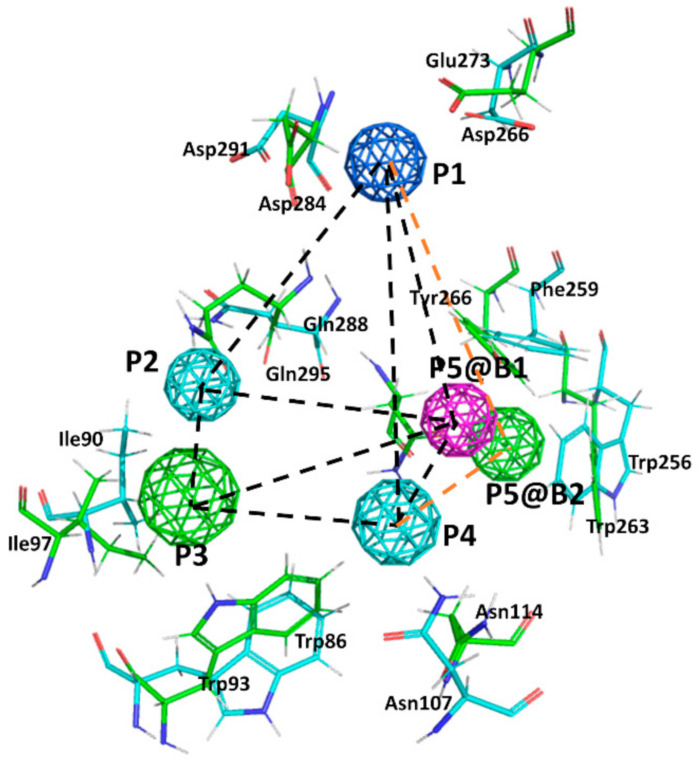
Figure 3.
Superposition of the B1 and B2 receptor pharmacophores. Colored spheres represent pharmacophore points (P1–P5) according to the following color code: dark blue represents a positive charge moiety; magenta a hydrogen bond accepting center; light blue a hydrogen bond donor/acceptor center; green an aromatic/lipophilic center. @B1 and @B2 is used to differentiate P5 for the B1 and B2 receptors, respectively. Consensus distances between common pharmacophore points are (black dotted lines): d(1,2) = 9 Å; d(1,3) = 14 Å; d(1,4) = 10.5 Å; d(2,3) = 6 Å; d(2,4) = 7 Å; d(3,4) = 7.5 Å. Specific distances for the B1 pharmacophore: d(1,5) = 9.5 Å; d(2,5) = 9.3 Å; d(3,5) = 9.5 Å; d(4,5) = 5.7 Å; whereas for the B2 pharmacophore are (orange dotted lines): d(1,5) = 11 Å; d(2,5) = 9 Å; d(3,5) = 8.8 Å; d(4,5) = 8.4 Å. Side chains of the main residues involved in defining the binding pocket for non-peptide ligands are explicitly depicted: green for the B1 and blue for the B2, respectively.

