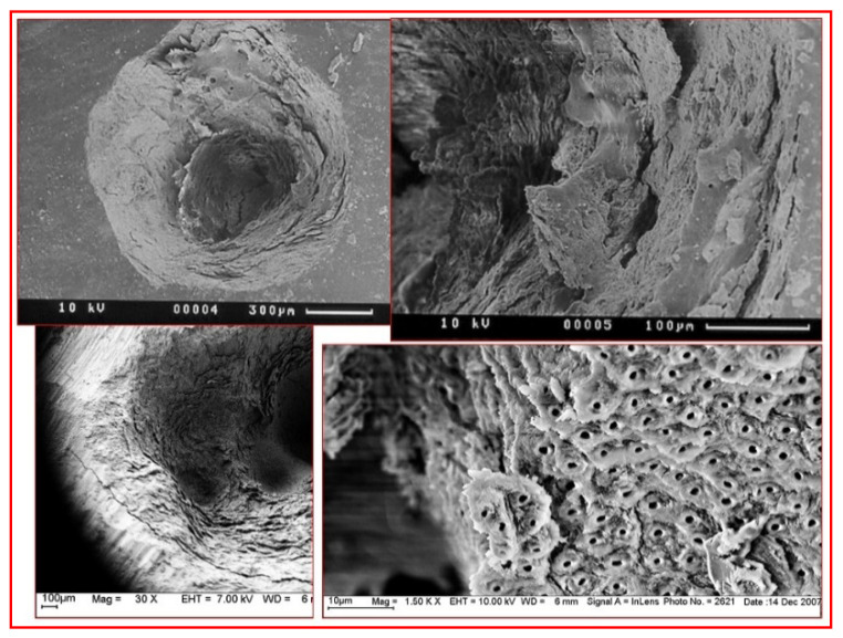
Figure 8.
Scanning Electron Microscope (SEM) micrographs of laser-mediated enamel and dentine ablation. Top left and right: the resultant enamel surface is rugged, fragmented and capable of accepting a resin-based composite restoration once any unstable fragments have been removed. Bottom left and right: dentine is rendered smear layer-free, with an intact and stable cut surface.

