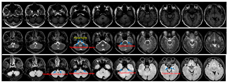
Figure 2.
Cranial nerves on clinical routine MRI. T1, T2, and Flair MRI images with a low spatial resolution (5-mm slice thickness in the axial plane with a 1-mm gap between slices) acquired by using a 3T MRI scanner are shown in the first, second, and third rows. Note that fewer cranial nerves can be seen in this figure than in Figure 1.

