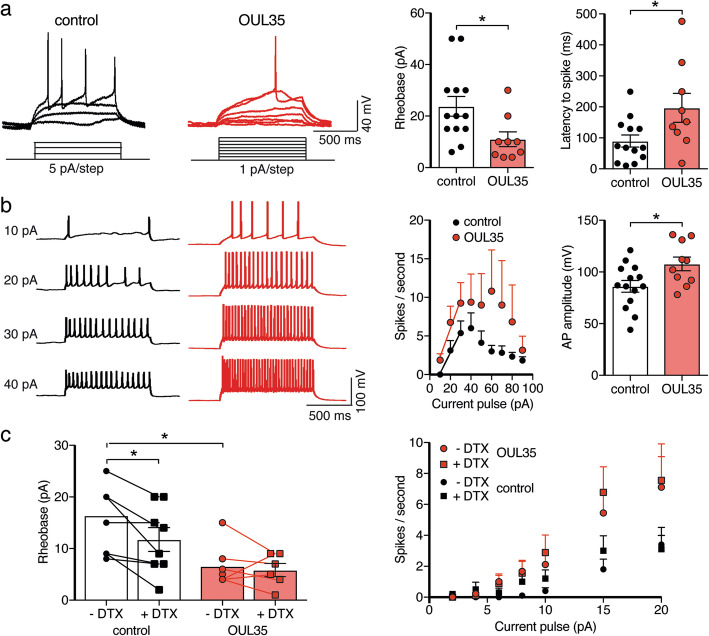
Fig. 6.
ARTD10 inhibition enhances excitability of hippocampal neurons via Kv1.1. a Left, representative current clamp recordings of APs elicited by step current pulses in control neurons and neurons treated with OUL35. Right, bar graphs represent the rheobase and the latency to the first spike. For cells with a RMP more positive than − 60 mV, the membrane potential was adjusted to ~ − 60 mV. b The number of spikes elicited by step current pulses were counted and for stimuli from 10 to 30 pA they were fitted with a linear function. Right, bar graphs summarize the AP amplitude from neurons with and without OUL35 treatment. c Left, bar graphs representing the rheobase. Right, summary of spikes/s with and without the Kv1 inhibitor α-dendrotoxin (DTX) and from neurons with and without OUL35 treatment. *p < 0.05 (Student’s t test)

