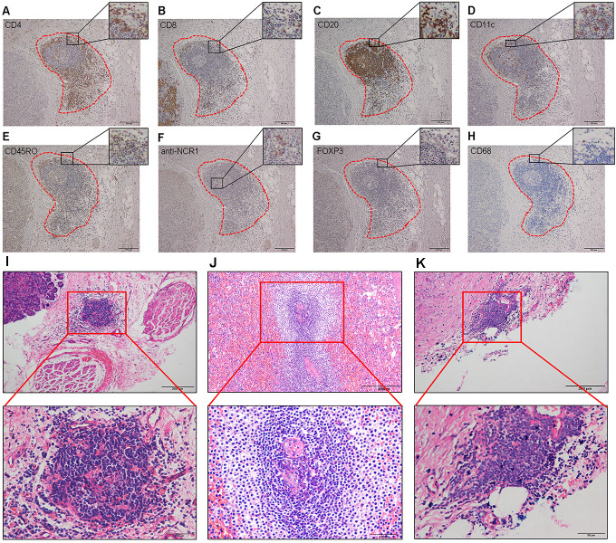
Figure 1.
Immunohistochemistry and histology of tertiary lymphoid structures (TLS). (A–H) Cellular composition of TLS. (A) CD4+ T cells. (B) CD8+ T cells. (C) CD20+ B cells. (D) CD11c+ dendritic cells. (E) CD45RO+ memory T cells. (F) anti-NCR1+ natural killer cells. (G) FOXP3+ regulatory T cells. (H) CD68+ tumor-associated macrophages. Magnification: 100×. (I–K) TLS stained with H&E. The presence of TLS in NF-PanNETs (I), functional PanNETs (J), and PanNECs (K). Top row, magnification: 100×. Bottom row, magnification: 400×. NF-PanNETs, non-functional PanNETs; PanNECs, pancreatic neuroendocrine carcinomas; PanNETs, pancreatic neuroendocrine tumors.

