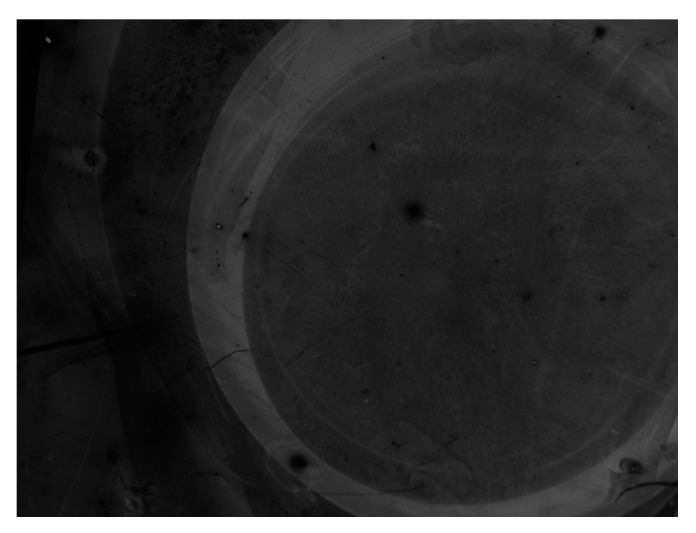
Figure A3.

Image by epifluorescence microscopy corresponding to the hybridization of the Sequence 2 probe with its complementary strand, performed 17 months after the probe grafting. The analysis was carried out on Si/SiO2 (100 nm) substrates functionalized by UV-assisted GOPS protocol, using an exposure time of 3 s.
