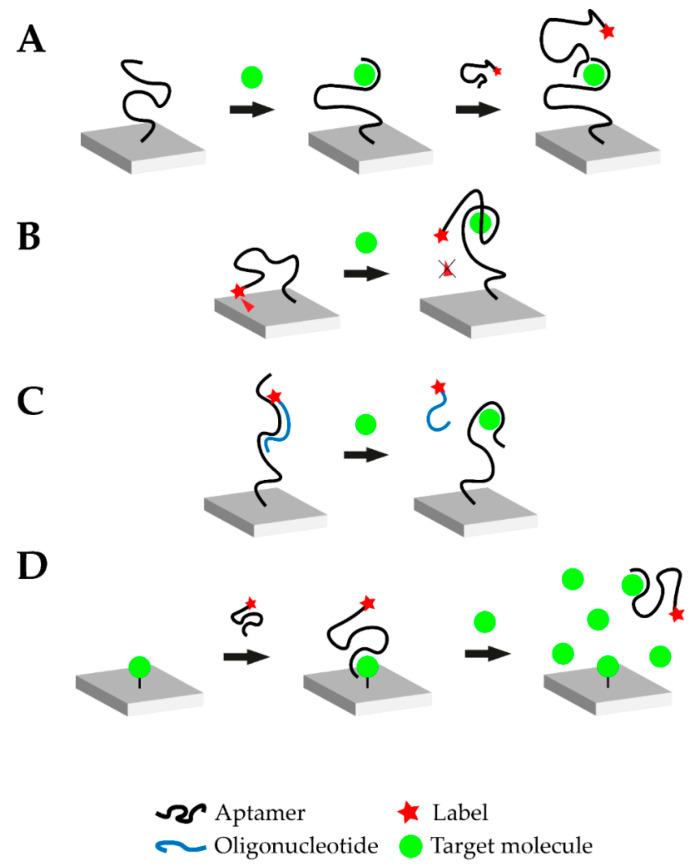
Figure 1.
Overview of the different possible operational modes of aptasensors. (A) Sandwich or sandwich-like mode: One aptamer is immobilized on a surface and the target molecule is added. Upon addition, the aptamer-target complex is formed. Afterwards, another labeled aptamer against the respective target is introduced which binds to a different epitope of the target molecule. Transduction is carried out via the labeled aptamer. (B) Target-induced structure switching (TISS) mode: The labeled aptamer is immobilized on a surface. In this example, a fluorescence molecule is linked to the aptamer and brought near a quenching surface. Upon target addition, the aptamer structure changes and the fluorescence molecule is no longer quenched. In this case, transduction is carried out via fluorescence measurement. (C) Target-induced dissociation (TID) mode: A complementary, labeled oligonucleotide hybridizes with the immobilized aptamer. Upon target addition, the aptamer-target complex is formed and the oligonucleotide is displaced. After washing steps, the respective signal decreases and transduction can be carried out. (D) Competitive replacement (CR) mode: In this example, the target molecule is immobilized on the surface. The labeled aptamer is added and forms the aptamer-target complex. When the target molecule is added in excess, the aptamer preferably binds to the free molecule and is removed from the immobilized target molecule. This change can then be monitored.

