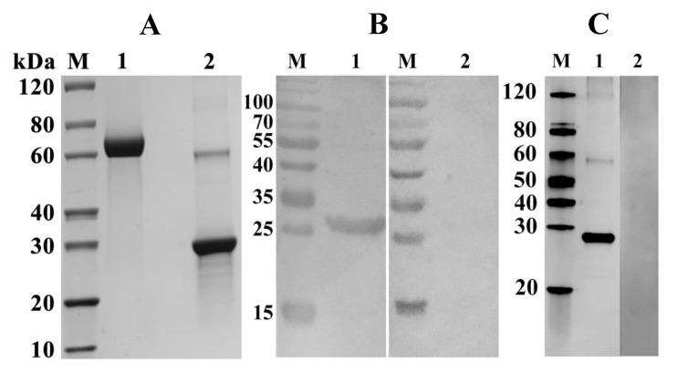
Figure 3.
SDS–PAGE and Western-blot analyses of the purified recombinant protein, and rFgTPx from the sonicated pellet of E. coli. (A) Proteins were resolved on 12% acrylamide gels and stained with Coomassie brilliant blue R250. Lane M: protein molecular weight marker in kDa; Lane 1: 5 μg bovine serum albumin; Lane 2: purified rFgTPx appeared as a single band of ~26 kDa. (B,C) The protein of interest was run under non–reducing conditions and visualized using a chemiluminescent horseradish peroxidase substrate. Lane M: protein molecular weight marker in kDa. (B) Lane 1 was loaded with rFgTPx. Serum from F. gigantica–infected goats detected a single band of ~26 kDa; Lane 2 was loaded with rFgTPx that did not react with serum of uninfected goat. (C) Lane 1 was loaded with rFgTPx and incubated with rabbit serum containing specific anti-rFgTPx antibodies, showing a single band at ~26 kDa band; Lane 2 was loaded with rFgTPx that did not react with naïve rabbit serum.

