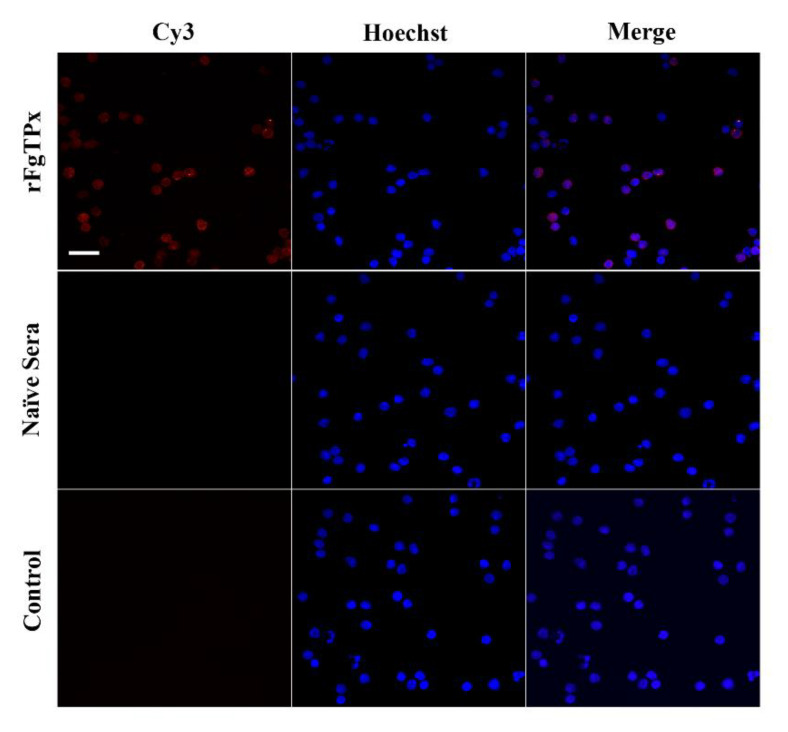
Figure 4.
Fasciola gigantica–derived rFgTPx binds to the surface of goat PBMCs. Visualization of rFgTPx attachment to PBMC surfaces was carried out by incubation of PBMCs treated or untreated with rFgTPx with rabbit anti–rFgTPx primary antibody. Hoechst (blue) and Cy3-conjugated secondary antibody (red) were used to stain host cell nuclei and rFgTPx, respectively. Positive staining of cell surface was detected in rFgTPx–treated cells only. No staining was detectable in control, untreated cells. Scale bars = 10 µm.

