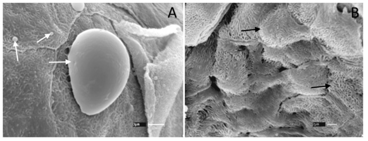
Figure 7.
Scanning electron microscopy (SEM) of the vaginal canal from BALB/c mice infected with 3 × 10⁵ yeasts of C. albicans ATCC 90028. (A) Control group, infected, and treated with neutral cream only. Arrow indicates the presence of C. albicans yeasts on the tissue. Scale bar = 3 µm (B) Section of vaginal canal from mice treated with 500 µM of limonene cream. Arrows point to the squamous cells present in the epithelium. Scale bar = 2 µm. Three samples per group were analyzed.

