FIGURE 2.
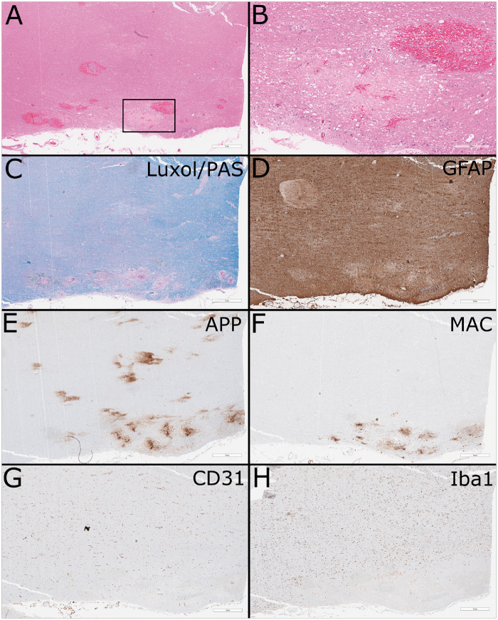
Routine histology and immunohistochemistry. (A) Low-power hematoxylin and eosin-stained formalin-fixed paraffin-embedded section of cerebellar peduncle showing areas of vacuolation and acute hemorrhage (scale bar: 1 mm). Areas of cerebellar gray matter were also focally involved (not shown). (B) Higher-power view of region of interest showing numerous axonal spheroids and minimal cellular reaction (scale bar: 300 µm). (C) Luxol fast blue/periodic acid-Schiff stain shows loss of myelin without evidence of active demyelination (scale bar: 1 mm). (D) GFAP stain shows loss of astrocytes in the lesion with moderate activation of the surrounding astrocytes (scale bar: 1 mm). (E) Amyloid precursor protein stain highlights the axonal damage (scale bar: 1 mm). (F) Membrane attack complex highlights vessels in and at the periphery of the lesion (scale bar: 1 mm). (G) CD31 shows widespread paucity of vessels in region of the peripheral lesion, with relative preservation in the central white matter (scale bar: 1 mm). (H) Iba1 stain shows near total loss of microglia in the peripheral lesion and surrounding parenchyma, in comparison to the relative normal density in the central white matter (scale bar: 1 mm).

