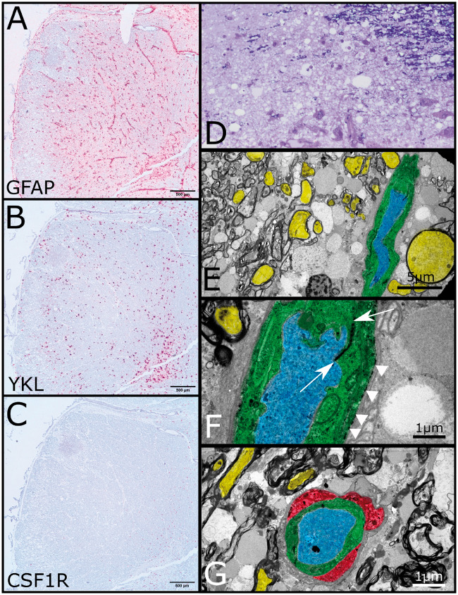
FIGURE 3.
In situ hybridization and electron microscopy. (A) In situ hybridization for GFAP of formalin-fixed paraffin-embedded embedded medulla demonstrating expected perivascular distribution and loss of expression in the vacuolated lesion (scale bar: 500 µm). (B) In situ hybridization demonstrates YKL expression in activated astrocytes (scale bar: 500 µm). (C) In situ hybridization for CSF1R fails to highlight any residual microglia (scale bar: 500 µm). (D) Thick section of plastic embedded brainstem stained with toluidine blue. Upper right corner shows lesion edge with preserved myelinated fibers the remaining tissue shows severe vacuolation with some residual cellular elements and vessels at bottom. (E) Low-power transmission electron micrograph from edge of lesion demonstrating axons (yellow) with retained surrounding compact myelin. Dilated axonal profiles with and without retained subcellular organelles are interspersed amongst the myelinated fibers. A single blood vessel courses from top to bottom on the right side. The endothelium is colored green, whereas the lumen is colored blue (scale bar: 5 µm). (F) Higher-power transmission electron micrograph of blood vessel showing electron dense junction between apposed endothelial processes (between arrows) and electron dense deposits along endothelial basement membrane (arrowheads) consistent with complement deposition (scale bar: 1 µm). (G) Rare perivascular microglial (red) present at the lesional edge shows cytoplasmic vacuolation with electron dense bodies (scale bar: 1 µm).

