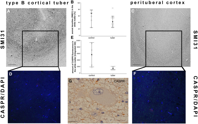
FIGURE 3.
Loss of nodes of Ranvier while the number of axons stay intact. (A) Cortex of a type B cortical tuber displaying the presence of phosphorylated neurofilament-positive axons (SMI31) at the gray-white matter border with the corresponding loss of CASPR-positive nodes of Ranvier (D; CASPR/DAPI). (B) No differences in SMI31-positive axons could be detected between the subgroups (white matter: p = 0.126; Mann-Whitney-U test). (C) Perilesional sample of the same patient as in a showing and normal number of axons (SMI31) and nodes of Ranvier (F; CASPR/DAPI) at the gray-white matter border. (E) Integrated optical density of CASPR fluorescence is reduced in cortical tubers (Mann-Whitney-U test, p = 0.016). Filled dots equal median, whiskers show 95% confidence intervals. (G) Abnormal arrangement of nodes of Ranvier in the presence of a giant cell. Scale bar in A and C equals 200 µm. Scale bar in e equals 25 µm. WM, white matter; GM, gray matter; CASPR, contactin-associated protein.

