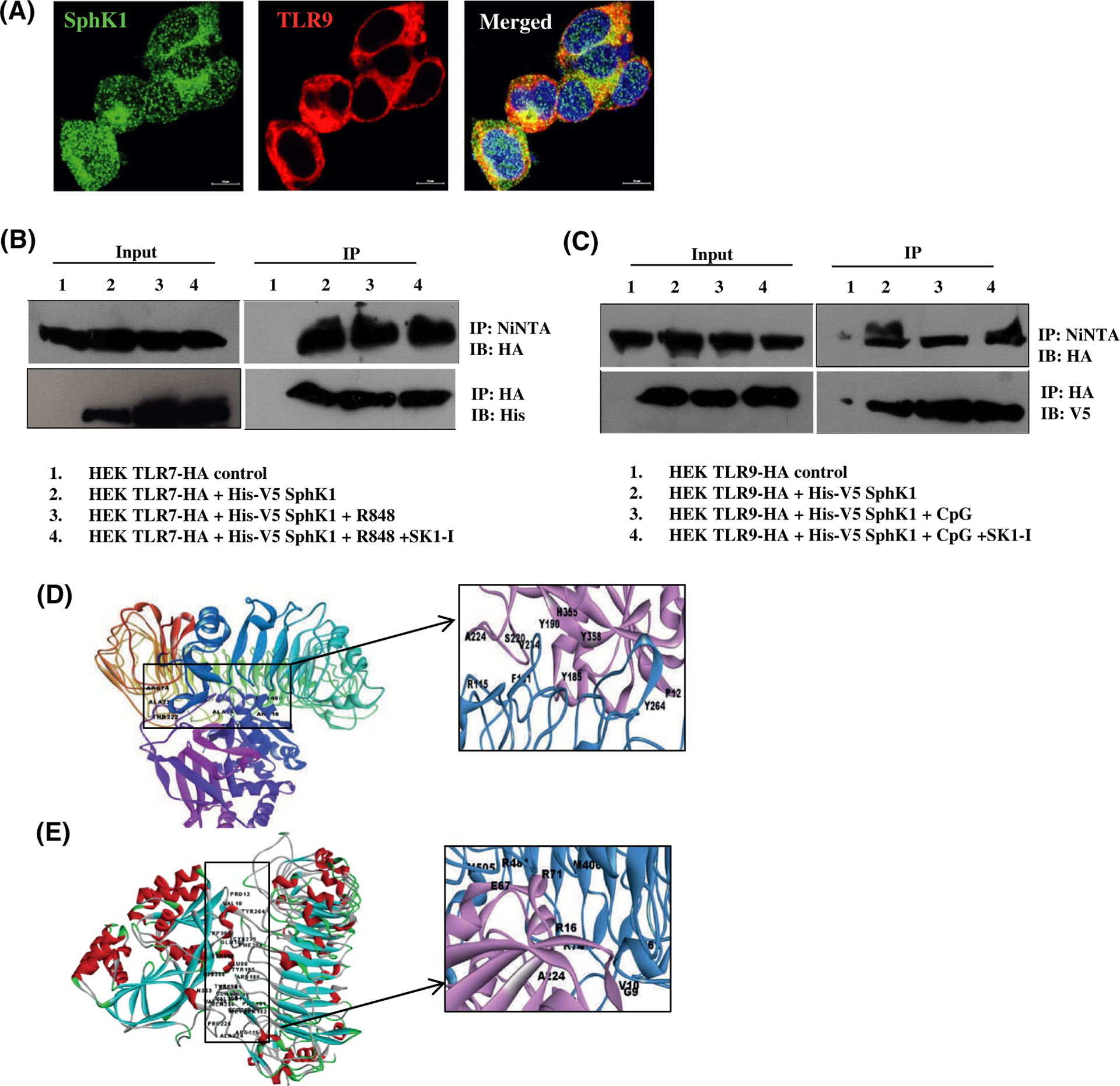
FIGURE 4.

SphK1 interacts with TLR7 and TLR9. A, HEK293 cells stably expressing HA-TLR9 transiently transfected with His-V5-SphK1. Representative images of immunofluorescence microscopy after staining with antibodies against TLR9 and SphK1. Images are representative of three independent experiments. Scale bar: 10 μm (B, C) HEK293 cells stably expressing HA-TLR7 (B) or HA-TLR9 (C) transiently transfected with His-V5-SphK1 and treated with R848, CpG-ODN or SK1-I as indicated. SphK1, TLR7, and TLR9 pulled down with Ni-NTA or HA affinity purification beads, separated by SDS-PAGE, and immunoblotted as indicated. Input exposed differently (D-E) Docked complexes of TLR9 and SphK1 (D) and TLR7 and SphK1 (E) obtained from ZDOCK server. The H-bond forming residues are marked in the inset (TLR9, TLR7- blue, SphK1- pink)
