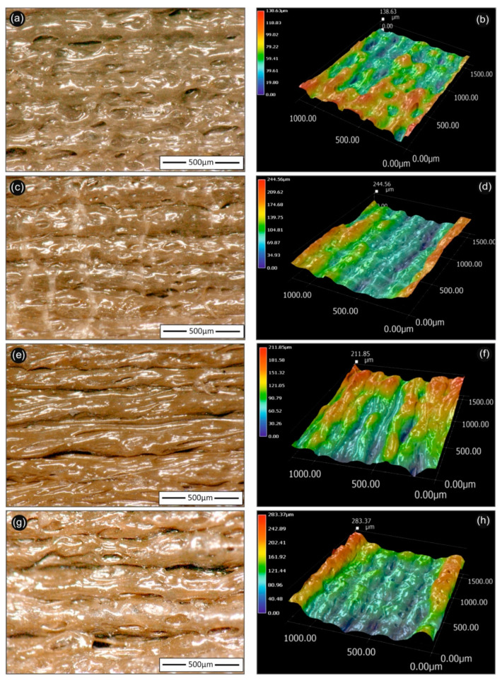
Figure 6.
Optical microscopy 2D (left hand-side) and 3D (right hand-side) images of 3D printed PLA/AgNP nanocomposites (at 100 µm printed layer thickness), utilizing PLA/Ag/PEG (rec-01)—(a,b); PLA/Ag/PEG (rec-02)—(c,d); (PLA/Ag/PVP (rec-03)—(e,f); and PLA/Ag/PVP (rec-04)—(g,h) extruded nanocomposite filaments, respectively. The 500 µm scale bar is inserted and shown in each figure in the left hand-side images, while the respective 3D images are shown in the right hand-side with the corresponding color scale bar indicating the micro-roughness of each sample.

