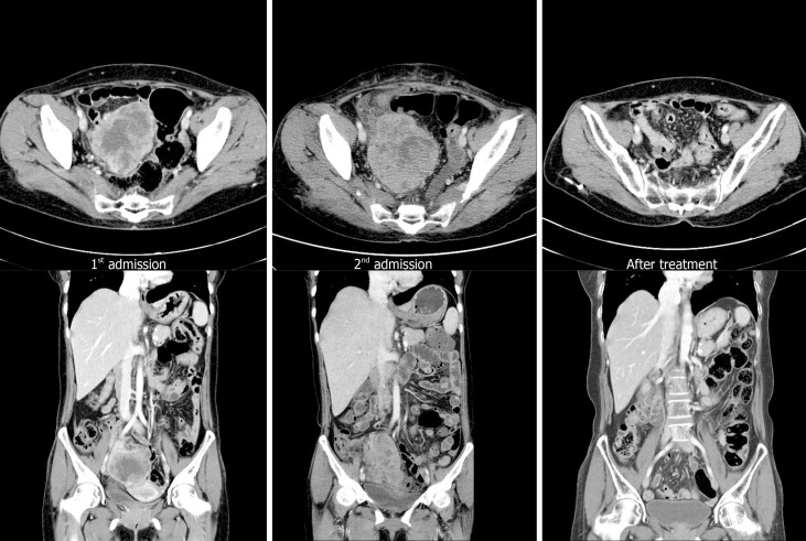
Figure 1.
Computed tomography at admission and follow-up. Computed tomography (CT) scan performed at first admission showing an 8 cm × 7 cm size mass only in the pelvic cavity. However, 6 wk after surgery (second admission), CT scan shows a mass that was larger than the initial mass in the pelvic cavity with peritoneal seeding and para-aortic lymphadenopathy (arrow). These lesions are almost no longer observed after chemotherapy.

