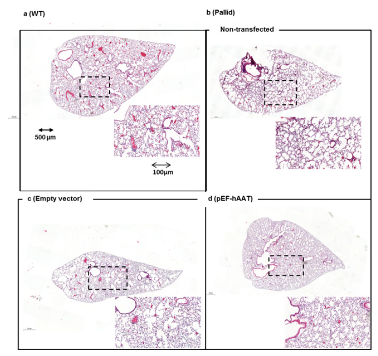
Figure 3.
Left lobe lung histology after intrahepatic AAT gene transfer by electroporation in vivo: Representative micrographs of the lungs of animals from different groups. (a) Normal lung from wildtype (WT) (C57BL/6J) mice, (b) air space enlargement visible in the non-transfected pallid mice and (c) in the animals treated with empty vector, (d) animals treated with the pEF-AAT plasmid: ×20 magnification, scale bar 500 µm. Inset, images at higher magnification as indicated by dashed frame (digital zoom; scale bar 200 µm).

