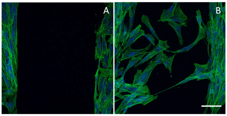
Figure 5.
CLSM (confocal laser scanning microscope) images of a cell substrate (normal human dermal fibroblasts) with a free cell area (lesion) obtained using an insert (panel (A)) and the same substrate 24 h after the insert removal characterized by cells migrated/proliferated in the free area (panel (B)) (the cell substrate has been stained with phalloidin-FITC (green, F-actin filaments) and DAPI (blue, nucleus)). Scale bar 100 μm. Modified from [135]. CC BY 4.0.

