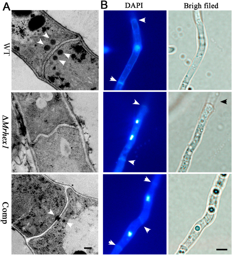
Figure 4.
Microscopic observations: (A) transmission electron microscope observation showing the presence or absence of Woronin bodies (arrowed) in the WT and mutant cells. Bar, 0.5 μm; (B) the co-staining of the mycelium cells for detecting nuclei and septa (arrowed). The broken end of the ΔMrHex1 mycelium is arrowed for its bright field image. Bar, 5 μm.

