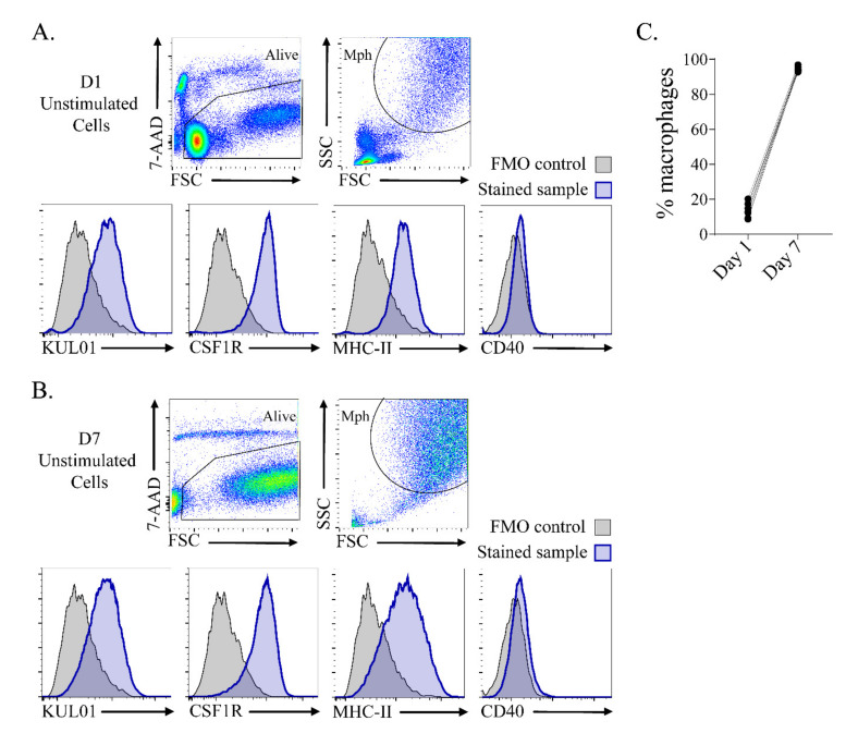
Figure 2.
Adherent cells differentiated to KUL01+ CSF1R+ MHC-II+ macrophages (Mph) after 7 days of culture. (A) Adherent cells were characterized after 24 h of culture by flow cytometry. (B) Adherent unstimulated cells were characterized after 7 days of culture by flow cytometry. The cells were selected for viability (7-AAD−), forward and side light scatter (FSC vs. SSC), and assessed for the expression of KUL01, CSF1R, MHC-II and CD40. The histograms show expression of the macrophage markers in blue and fluorescent-minus-one (FMO) staining controls in grey. (C) The percentages of macrophages from day 1 and day 7.

